Research
- Resources
- Welsh Lab
- McCarthy Lab
In mammals, circadian (ca. 24h) clocks in the brain and throughout the body orchestrate daily patterns of physiology and behavior. These daily patterns persist under constant conditions as "circadian rhythms". The Welsh lab studies circadian rhythms in cells using bioluminescence imaging to monitor clock gene expression. We are interested in the autonomy, heterogeneity, and coupling of cellular circadian clocks, particularly the "master" clock cells of the brain, the neurons of the suprachiasmatic nucleus. We are also interested in how defects in these mechanisms may contribute to sleep and circadian rhythm disorders in humans, including mood disorders.
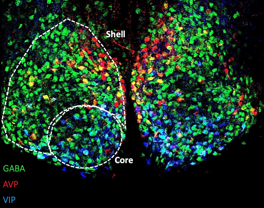
In collaboration with the Dulcis Lab , Welsh Lab members are studying the effects of photoperiod on protein and gene expression within the mammalian suprachiasmatic nucleus (SCN). Pictured below is a coronal view of the SCN stained for arginine vasopressin (red), GABA (green) and vasoactive intestinal peptide (blue), three proteins involved in the integration and communication of light signals.